Figure 5.
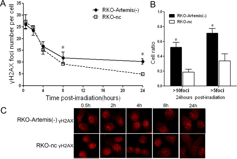
Impaired double-strand break (DSB) rejoining in RKO-Artemis (−) cells after IR. (A) The average number of γ-H2AX foci per cell was counted and shown. Each bar represents mean ± SD. At least 100 nuclei were evaluated per sample. (B) Cell ratio with residual foci more than 10 or 5 at 24 h after IR. Data represent the results from three independent experiments. Error bars represent SD of three independent experiments. *Statistical significance (p < 0.05). (C) Representative image of γ-H2AX foci under an immunofluorescence microscope.

