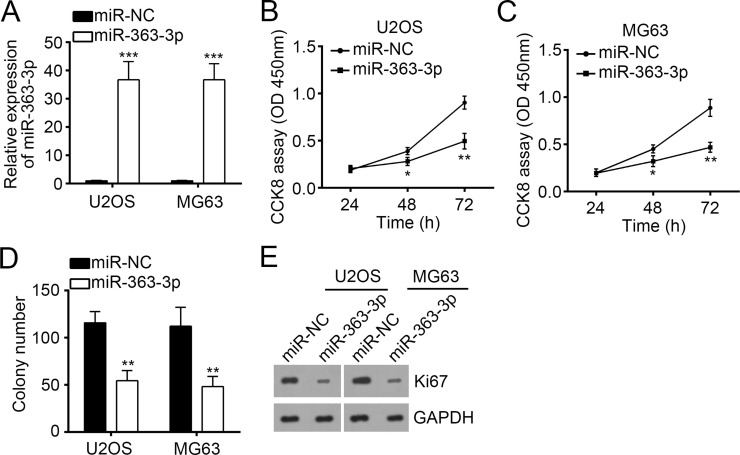
Figure 2.
Overexpression of miR-363-3p inhibited cellular proliferation of OS cells. (A) Reverse transcription and quantitative real-time PCR (RT-qPCR) analysis of miR-363-3p expression in U2OS and MG63 cells transfected with miR-363-3p mimic or miR-NC. (B, C) Cell proliferation curves in U2OS and MG63 cells transfected with miR-363-3p mimic or miR-NC by cell counting kit-8 (CCK-8) assay. (D) Cell proliferation ability was assessed by colony formation assay with U2OS and MG63 cells transfected with miR-363-3p mimic or miR-NC. (E) Western blot analysis of Ki-67 expression in U2OS and MG63 cells transfected with miR-363-3p mimic or miR-NC. GAPDH was used as control. All data are representative of three independent experiments and expressed as mean ± SD. *p < 0.05, **p < 0.01, and ***p < 0.001.

