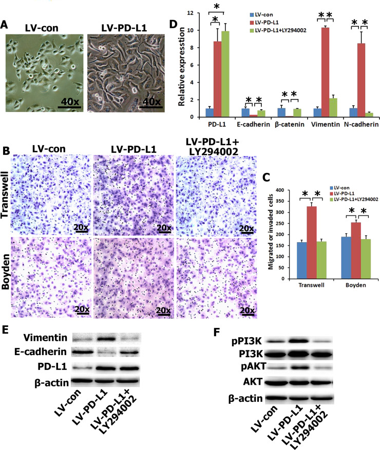
Figure 2.
Enhanced cell migration and invasion in PD-L1-expressing CNE2 cells via the PI3K/AKT pathway. (A) CNE2 cells coupled with an alteration of cellular morphology. (B, C) The migration and invasion were analyzed with an in vitro migration assay using a Transwell chamber and an in vitro invasion assay using a Matrigel-coated Boyden chamber, respectively. PI3K inhibitor (LY294002) abolished the migration and invasion capability induced by PD-L1. Representative photomicrograph is presented. The migrated cells were plotted as the average number of cells per field of view from five different experiments, as described in Materials and Methods. (D, E) The mRNA and protein expressions of EMT-related genes were detected by quantitative real-time (qRT)-PCR and Western blot with PD-L1-overexpression in CNE2 cells and pretreated with LY294002 (20 μM). (F) The protein expressions of PI3K/AKT signal pathway were detected by Western blot. *p < 0.05 compared to control.

