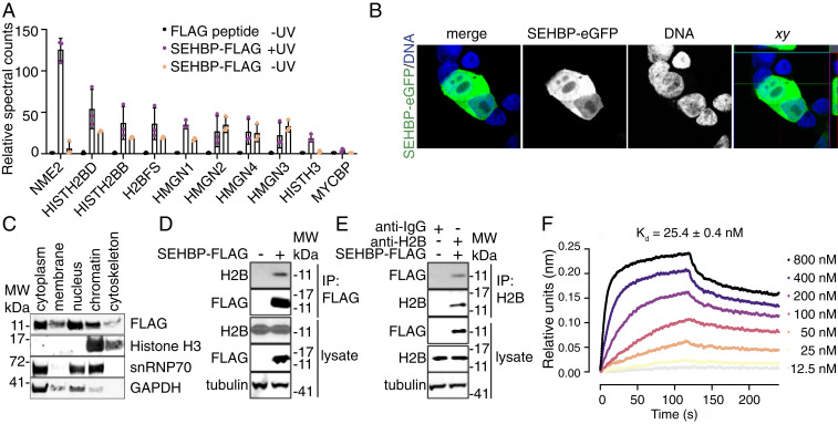
Fig. 3.
SEHBP interacts with Histone H2B in cells. (A) Relative spectral counts of the indicated anti-FLAG immunoprecipitated proteins after 48-h expression of SEHBP-AbK-FLAG in HEK293T cells followed by exposure to UV (n = 3; mean ± SD). (B) Representative confocal microscopy-derived images of SEHBP-eGFP localization (green) from HEK293T cells after 48 h of SEHBP-eGFP expression followed by fixation and exposure to Hoechst 33342. (C) Representative Western blotting analysis for the indicated proteins after 48-h expression of SEHBP-FLAG followed by subcellular fractionization. (D) Western blotting for endogenous H2B content after anti-FLAG immunoprecipitation from HEK293T cells expressing SEHBP-FLAG. (E) Western blotting analysis for SEBHP-FLAG content after anti-H2B immunoprecipitation from HEK293T cells expressing SEHBP-FLAG. (F) Sensorgram plot from biolayer interferometry experiments demonstrating the interaction between recombinant H2B and immobilized SEHBP.

