Fig. 1.
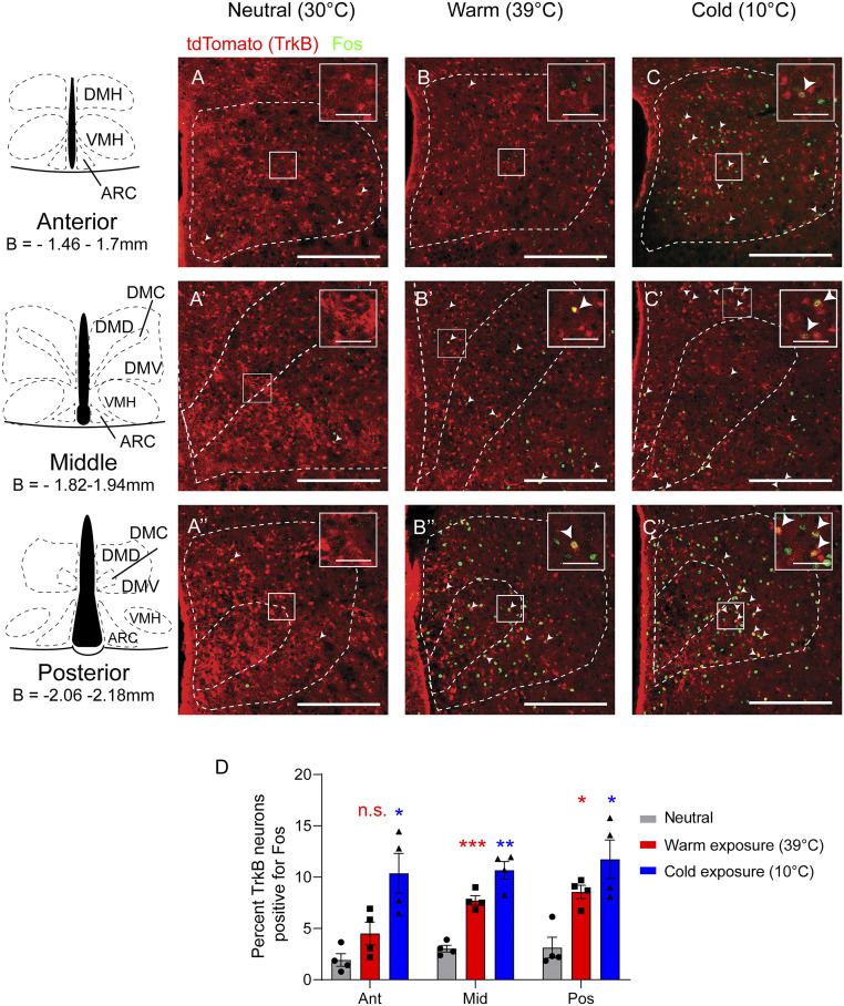
DMHTrkB neurons are sensitive to changes in environmental temperature. Representative images of Fos staining (green) in the DMH of Ntrk2CreER/+;Ai9 reporter mice after exposure for 2 h to (A–A”) thermoneutral (30 °C), (B–B”) warm (39 °C), or (C–C”) cold (10 °C) temperatures. tdTomato (red) marks TrkB-expressing cells in reporter mice. (Scale bars, 250 µm; inset scale bars, 50 µm.) (D) Quantification of Fos induction in TrkB-expressing neurons in the anterior (Ant), middle (Mid), and posterior (Pos) DMH after mice were exposed to different temperatures. Two-way ANOVA: temperature, F(2, 9) = 18.68; P = 0.0006; n = 4 mice per condition; Dunnett’s posttest versus neutral; n.s. = not significant, *P < 0.05, **P < 0.01, and ***P < 0.001. Values represent mean ± SEM. DMH, dorsomedial hypothalamus; DMD, DMH dorsal division; DMC, DMH central division; DMV, DMH ventral division; VMH, ventromedial hypothalamus; ARC, arcuate nucleus; B, bregma.

