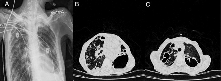
Figure 1.

A 65‐year‐old‐woman with Mycobacterium simiae respiratory infection. (A) Multiple scattered nodules were seen in both lung fields, especially the right with upper and middle preference. Opacity image was seen with the formation of a fluid surface in the lower zone of the left lung, which indicates a cavitary lesion. (B, C) Hydropneumothorax at the base of the left lung. Multiple nodules in lung tissue with several cavitary foci were observed in the right and left lung fields. A scattered consolidation patch was also evident in the lung field.
