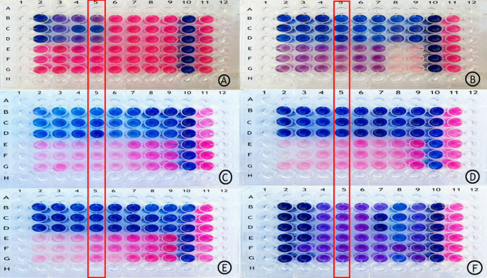
Figure 2.

Antibiotic resistance pattern using manual method (broth microdilution) using resazurin reagent: rows B, C, and D in all plates are related to standard Mycobacterium simiae (JCM12377) and rows E, F, and G in all plates are related to the clinical isolation of M. simiae. To ensure the accuracy of the results of each antibiotic for standard sample and clinical isolation, it was performed three times. The concentration of antibiotics was reduced from column 2 to column 9. Plate A contains the antibiotic moxifloxacin (0.25–32 μg/mL). Plate B contains the antibiotic clofazimine (0.015–2 μg/mL). Plate C contains the antibiotic streptomycin (0.5–64 μg/mL). Plate D contains the antibiotic clarithromycin (2–256 μg/mL). Plate E contains the antibiotic cotrimoxazole (0.03–4 μg/mL). Plate F contains the antibiotic amikacin (0.5–64 μg/mL). Columns 10 and 11 represent sterility control and growth control of each plate, respectively. In columns 1 and 12 and rows A and H, sterile distilled water is added to prevent the plates from drying out.
