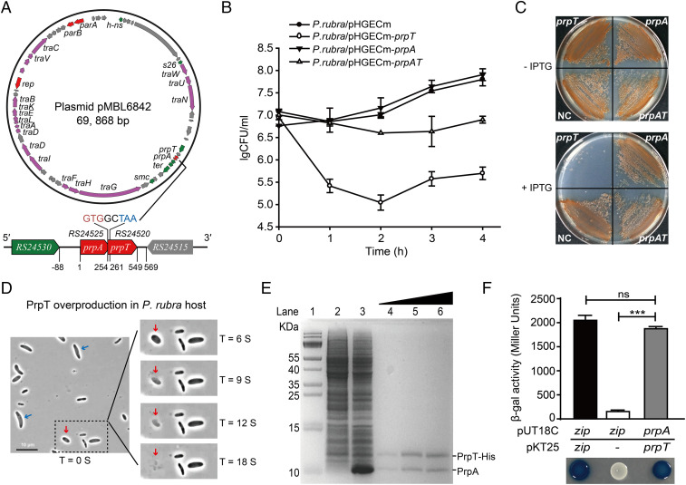
Fig. 2.
PrpT and PrpA constitute a TA pair. (A) Circular map of pMBL6842 and the position of the prpA-prpT operon. (B and C) Viability of cells overexpressing prpT, prpA, and prpA-prpT in P. rubra. IPTG, isopropyl β-ᴅ-thiogalactoside. (D) Morphologies of cells overexpressing prpT with 0.5 mM IPTG for 2 h over time (see Movie S1). The ghost and lysed cells are marked with blue and red arrows. (E) PrpT and PrpA form a complex in vitro. His-tagged PrpT and untagged PrpA were coproduced via pET28b-prpA-prpT-His (lane 3) and copurified with increasing concentration of imidazole (lanes 4–6). Lane 1: size marker; lane 2: NC (no IPTG). (F) The BACTH assay showed that PrpT interacts with PrpA. The data are from three independent cultures. SDs are shown, and statistical significance (NS, no significant; *P < 0.05; **P < 0.01; ***P < 0.0001) is indicated with asterisks in Figs. 2–4 and 6. Images shown in Figs. 2–6 are representative images.

