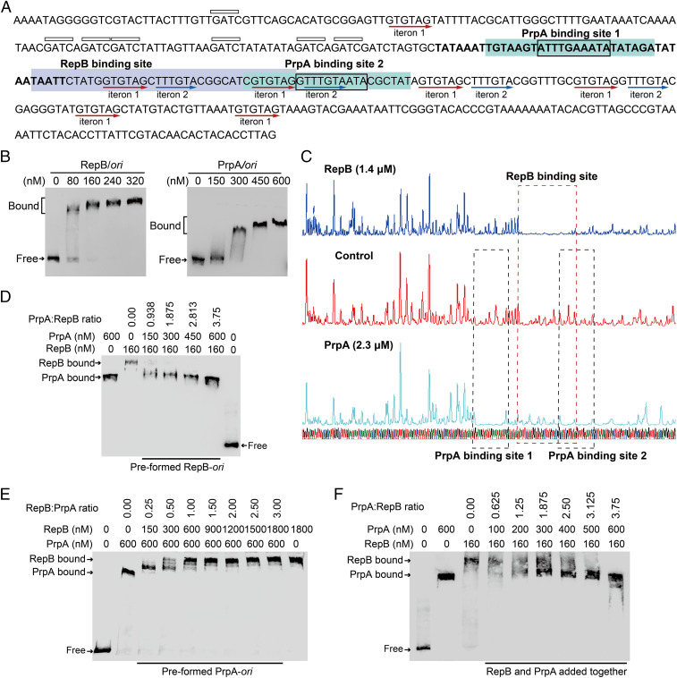
Fig. 5.
PrpA competes with RepB for binding to the pMBL6842 ori. (A) Nucleotide sequence of the pMBL6842 ori region. The AT-rich region is marked in bold letters. The 5′-GATC sites are indicated by boxes above the sequence. The iterons are underlined using red arrows. The binding sites of RepB and PrpA are highlighted with blue and green, respectively. The conserved binding motif of PrpA in site 1 and 2 is indicated by a box in the sequences. (B) EMSA results showing that RepB and antitoxin PrpA bind and shift the pMBL6842 ori. (C) DNase I footprinting assays used to determine the binding sites of RepB and PrpA. EMSA results showing that PrpA and RepB compete for binding to ori when the two proteins are added sequentially (D and E) or added at the same time (F).

