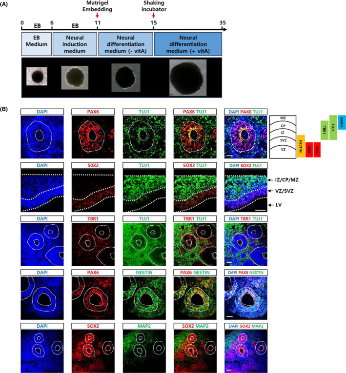
FIGURE 1.

Generation of cerebral organoids. A, Schedule of brain organoid generation using an unguided method. 10 The shape and relative sizes of brain organoids at each step were shown at the bottom. B, Brain organoids at 6 weeks of culture were sectioned and analysed for the expression of various neural markers: Neural progenitor markers (PAX6, Nestin and SOX2), early neuronal markers (TUJ1), mature neuronal marker (MAP2) and postmitotic cortical neuronal marker (TBR1) were shown. Scale bar, 50 μm. Layers and representative markers of human cerebral cortex were shown on the top & right side. CP, cortical plate; IZ, intermediate zone; LV, lateral ventricles; MZ, marginal zone; SVZ, subventricular zone; VZ, ventricular zone
