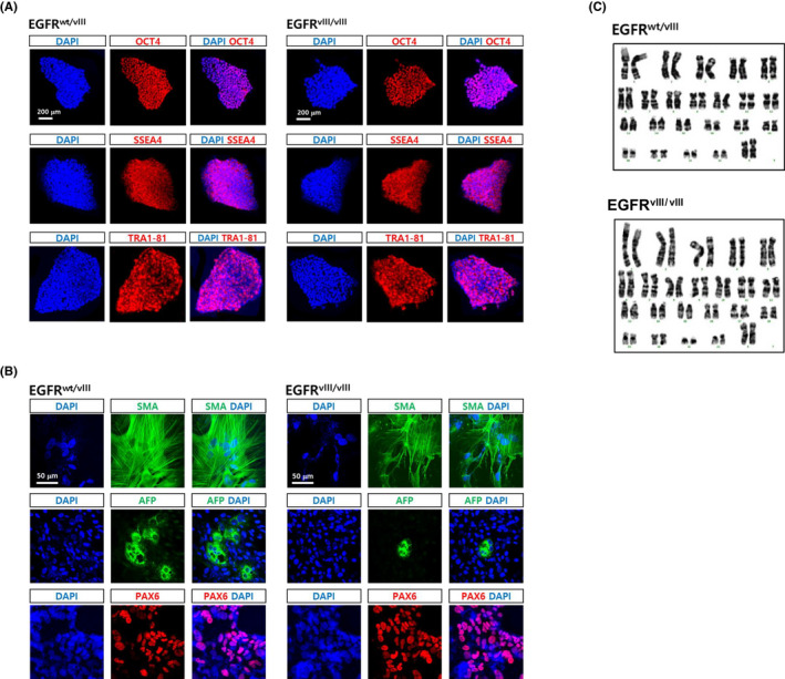
FIGURE 3.

Characterization of EGFRvIII‐hESC clones. A, Both monoallelic and biallelic EGFRvIII clones were immunostained with antibodies recognizing typical hESC markers, Oct4, SSEA4, Oct4 and Tra1‐81. DAPI staining (blue) were also performed to expose the presence of cells. Scale bar, 200 μm. B, The EGFRvIII clones were spontaneously differentiated into derivatives of the three germ layers for 3 weeks via EB formation. Smooth muscle actin (SMA), alpha‐fetoprotein (AFP) and PAX6 were detected by immunostaining as markers for mesoderm, endoderm and ectoderm, respectively. Scale bar, 50 μm. C, G‐banding analysis of the EGFRvIII clones was performed to examine the presence of gross chromosomal abnormality
