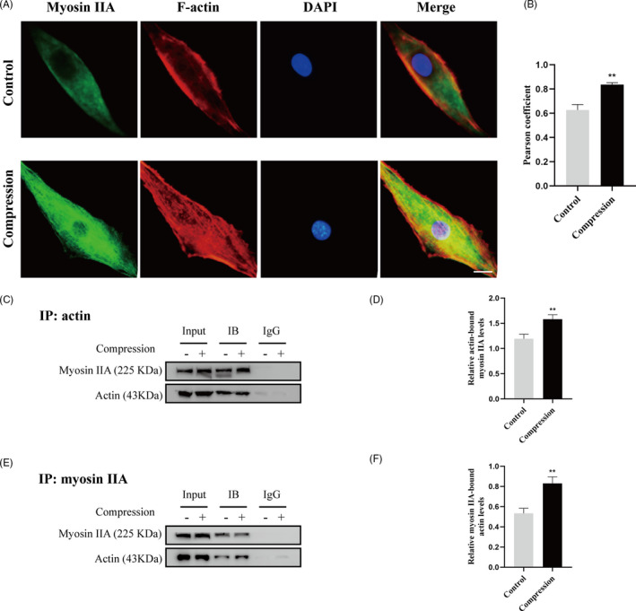
FIGURE 2.

Compression stress increased the interaction of myosin IIA with actin. (A) Human NP cells untreated or treated with compression stress for 36 h were stained with myosin IIA (green), F‐actin (red) and DAPI (blue). Scale bar: 5 μm. (B) The co‐localization of myosin IIA with F‐actin was evaluated on the basis of Pearson coefficients. (C, D) Protein interaction between myosin IIA and actin was determined by co‐immunoprecipitation. Following treatment, cell lysates were immunoprecipitated with anti‐actin antibody. Isotype‐matched (IgG) served as negative control. Each precipitated sample was detected for the presence of myosin IIA and actin by immunoblot analysis using specific antibodies. Whole cell lysates prior to the immunoprecipitation served as input controls. (E, F) Cell lysates were immunoprecipitated with anti‐non‐muscle myosin IIA antibody, the next steps are described above. Data were presented as the mean ± SD (n = 3). **P < .05, vs control
