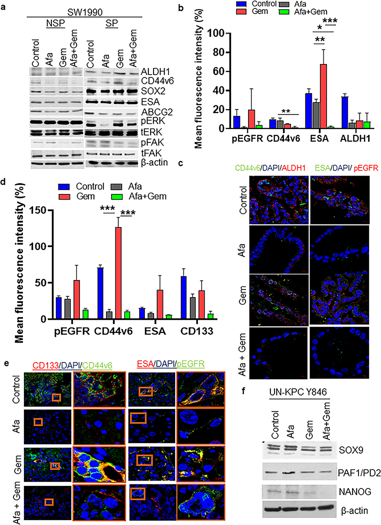
Fig. 5. Afatinib inhibited CSC stemness by downregulating multiple CSC markers in vitro and in vivo xenograft PC models.
a NSP and SP cells were isolated from SW1990 PC cells and subjected to drug treatment for 24 h. Immunoblot analysis of CSC (ALDH1, CD44v6, SOX2, ESA, and ABCG2), oncogenic/proliferative (total and activated ERK) and migratory (total and activated FAK) effectors upon afatinib treatment. (b - e) Co-immunolocalization analysis of CSC proteins in KPC tumoroids and PC xenograft tissues. Bar graph demonstrating quantification of mean fluorescent intensities of CSC proteins (pEGFR, CD44v6, ESA, ALDH1, and CD133) in KPC organoids (b) and PC xenograft tissues (N=5/group, *P<0.05, **P<0.01, ***P<0.001) (d). Representative confocal microscopic images are showing a response of CSC markers upon afatinib/gemcitabine treatment in KPC tumoroids (c) and xenograft tissues (e), scale bars=20 μM. (f) Western blot analysis of CSC markers (SOX9, PAF1/PD2, and NANOG) in mouse UN-KPC Y846 PC cells treated with/without afatinib and gemcitabine and combination. Beta-actin served as loading control in NSP-SW1990, SP-SW1990, and UN-KPC Y846 PC cells.

