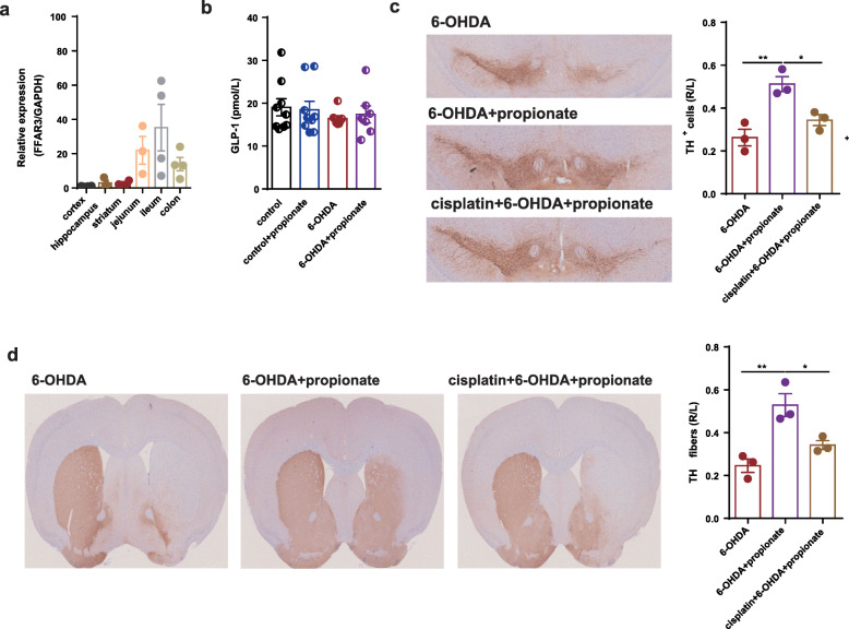
Fig. 8.
Enteric nervous system mediated the neuroprotective effects of propionate in 6-OHDA-induced PD mice. a The relative expression of FFAR3 in cortex, hippocampus, striatum, jejunum, ileum, and colon. n = 4. b Bar plot of the serum level of Glp-1 among different groups. n = 7–12. c (Left panel) Representative immunostaining showing TH-positive neurons in the SN. (Right panel) The average number of TH-positive neurons in the ST. n = 3 per group, 3 sections per mouse. d (Left panel) Representative immunostaining showing TH-positive fibers in the striatum. (Right panel) The quantification of TH-positive fibers in the striatum. Immunostaining: n = 3 per group, 3 sections per mouse. The data represent the mean ± SEM, p < 0.05 was set as the threshold for significance by one-way ANOVA followed by post hoc comparisons using Tukey’s test for multiple groups’ comparisons, *p < 0.05, **p < 0.01, ***p < 0.001, ****p < 0.0001

