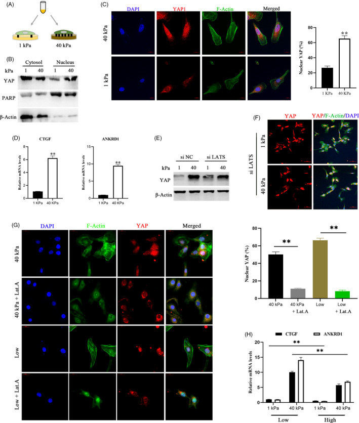
FIGURE 3.

YAP is regulated by ECM stiffness independent of the Hippo pathway and requires tension of the actin cytoskeleton. a. Schematic representation of bEECs plated on hydrogels with different rigidities (40/1 kPa). B, Western blotting for YAP in nuclear and cytoplasmic protein fractions from bEECs plated on 40 kPa and 1 kPa fibronectin‐coated hydrogels for 48 hour, n = 3. C, Confocal immunofluorescence images (left, n = 2) and quantifications of nuclear and cytoplasmic subcellular localization (right, n = 15) of YAP in bEECs plated on hydrogels with different rigidities. Scale bars, 10 μm. D, RT‐ qPCR analysis of mRNA levels of CTGF and ANKRD1 in bEECs plated on hydrogels with different rigidities, n = 3. E, Protein expression of YAP in bEECs transfected with siNC or siLATS at 40/1 kPa hydrogels, n = 3. F, Representative immunofluorescence of YAP in bEECs transfected with si LATS and cultured on hydrogels, n = 2. Scale bars, 50 μm. G, Confocal immunofluorescence images (left, n = 2) and quantification of nuclear and cytoplasmic subcellular localization (right, n = 15) of YAP in bEECs plated at 40 kPa or low density. Cells were also treated with the F‐actin inhibitor latrunculin A (Lat.A, 0.5 μM) or PBS (control) for 24 hour. Scale bars, 20 μm. h. RT–qPCR of bEECs grown under low or high conditions on the indicated hydrogels, n = 3. Experiments were repeated n times with two biological replicates. Data are shown as the mean ± SEM. P values were determined by an unpaired two‐sided t test (c, d, g) and two‐way ANOVA (h). **P < .001. DAPI, blue, nuclei; F‐actin, green, cell boundaries; YAP, red. See also Figure S2d–h
