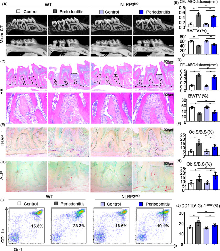
FIGURE 1.

NLRP3 deficiency protects against alveolar bone loss in ligature‐induced periodontitis. Two‐month‐old NLRP3KO mice and their WT littermates were used. 5‐0 silk ligature was tied around the right maxillary second molar and the other side was left untied to serve as the control group. All the mice were sacrificed 10 d after placement of the ligature. (A) Representative three‐dimensional (3D) micro‐CT scanning images (upper panels) and reconstructed sections (lower panels) along the longitudinal direction of the maxillae. (B) The distance of the cemento‐enamel junction (CEJ) to the alveolar bone crest (ABC) in mm and the percentage of BV/TV were analysed. BV/TV, which represents bone volume fraction, stands for Bone Volume over Tissue Volume. (C) Representative images of H&E‐stained paraffin sections. (D) The distance from the CEJ to ABC in mm and BV/TV (%) were determined. (E) Representative images of TRAP‐stained paraffin sections. (F) The percentage of alveolar bone surface covered by TRAP‐positive osteoclasts (Oc.S/B.S) was determined by histomorphological analysis. TRAP‐positive osteoclast surface (Oc.S/B.S) refers to the percentage of alveolar bone surface covered by mono‐ and multinucleated osteoclasts (ideally identified as TRAP‐positive). (G) Representative images of ALP‐stained paraffin sections. (H) The percentage of alveolar bone surface covered by osteoblasts (Ob.S/B.S) was determined. (I) Cells from periodontal tissues were collected and analysed by flow cytometry. Representative dot‐plot shows the population of CD11b+Gr‐1−/low cells. (J) The percentage of CD11b+Gr‐1−/low cells in the periodontal tissues was determined. All error bars represent SD, N = 7. One‐way ANOVA followed by Dunnett's post‐hoc multiple comparisons was performed. *P < .05 in the indicated groups
