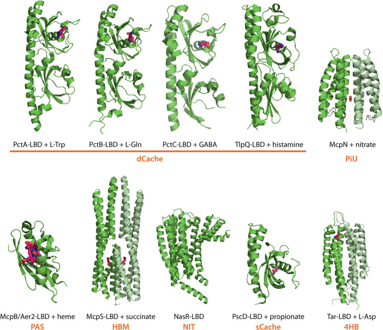
FIG 4.
Diversity of P. aeruginosa chemoreceptor LBDs. 3D structures of LBDs from PctA (PDB ID 5T7M), PctB (5LTO), PctC (5LTV) (38), TlpQ (6FU4) (39), McpN (6GCV) (46), and McpB/Aer2 (4HI4) (88) are shown (all P. aeruginosa). For the remaining protein families (Fig. 3), the structures of homologous domains from other species are shown, namely, P. putida McpS (HBM domain, 2YFB) (59), Klebsiella oxytoca NasR (NIT domain, 4AKK) (196), P. syringae PscD (sCache domain, 5G4Z) (197), and Salmonella enterica serovar Typhimurium Tar (4HB domain, 2LIG) (198). Bound ligands are shown in stick mode, and the LBD type is shown in orange. The monomers of LBD dimers are shown in different shades of green.

