Figure 5.

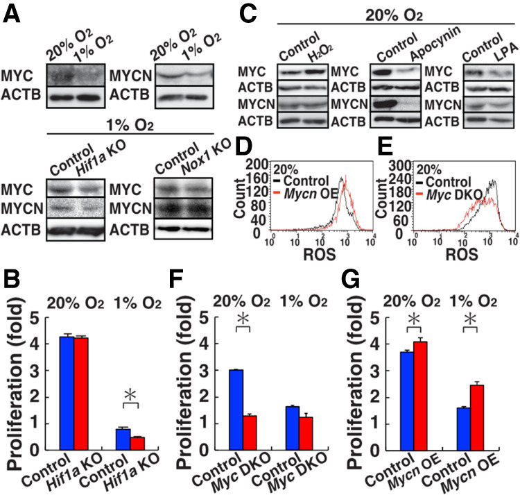
Induction of MYC/MYCN by ROS. (A) Western blot analysis of MYC/MYCN in WT (20% and 1% O2), Hif1a KO (1% O2), and Nox1 KO (1% O2) GS cells (n = 3). (B) Proliferation of Hif1a KO GS cells (n = 3). Cell recovery was determined after culturing under hypoxia for 3 d. (C) Western blot analysis of MYC/MYCN in WT GS cells after exposure to H2O2, apocynin or LPA (n = 3). (D) Flow cytometric analysis of ROS levels by CellROX Deep Red after Mycn overexpression in WT GS cells 11 d after transfection (n = 3). (E) Flow cytometric analysis of ROS levels in Myc DKO GS cells by CellROX Deep Red. (F) Proliferation of Myc DKO GS cells (n = 3). Cells were recovered after 4 d. (G) Proliferation of Myc DKO GS cells transfected with Mycn (n = 3). Cells were recovered 8 d after transfection. Asterisk indicates statistical significance (P < 0.05).
