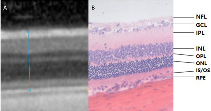
Figure 3. Corresponding relationship between OCT examination result and pathological section of rat retina (the range of blue arrow indicates total retinal thickness, HE *400).
(A) The OCT examination result of rat retina. (B) A pathological section of the retina of rats. OCT images and pathological findings of each layer of the retina can be seen in the two images. Blue arrows indicate the total retinal thickness. NFL: Nerve Fiber Layer; GCL: Ganglion Cell Layer; IPL: Inner Plexiform Layer; INL: Inner Nuclear Layer; OPL: Outer Plexiform layer; ONL: Outer Nuclear layer; IS/OS: Inner Segment/Outer Segment; RPE: Retinal Pigment Epithelium.

