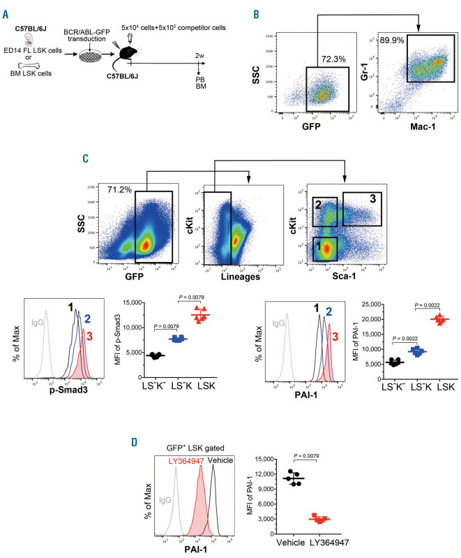
Figure 1.
TGF-−iPAI-1 signaling is activated in chronic myeloid leukemia - leukemic stem cells. (A) Schema for experiments. (B) Representative flow cytometric profiles of contribution of BCR/ABL-GFP+ cells to the myeloid (Mac-1+/Gr-1+) peripheral blood (PB) output post transplantation. (C) Representative flow cytometric profiles and mean fluorescent intensity (MFI) (n=6) for p-Smad3 and intracellular plasminogen activator inhibitor-1 (iPAI-1) expressions in freshly isolated BCR/ABL-GFP+ immature chronic myeloid leukemia (CML) cells in the bone marrow (BM). MFI of LSK: Lin–c-kit+Sca-1+; LS–K: Lin–c-kit+Sca–1–; LS–K–: Lin–c-kit–Sca-1–. (D) Representative flow cytometric profile and MFI for iPAI-1 expression in freshly isolated BCR/ABL-GFP+ immature CML cells in the BM of vehicle- or LY364947-treated mice (n=5). Data represent means ± standard deviation. Statistical significance was determined by Mann-Whitney unpaired t-test. P<0.001, by a Kruskal-Wallis test. TGF-: transforming growth factor-; BCR: breakpoint cluster region; ABL: Abelson kinase; GFP: green florescent protein; SSC: side scatter.

