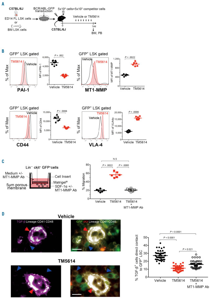
Figure 5.
iPAI-1 blockade increases membrane type-1 metalloproteasedependent cellular motility. (A) Schema for experiments. (B) Representative flow cytometric profiles and mean fluorescent intensity (MFI) (n=6-9) for iPAI-1, MT1-MMP, CD44, and VLA-4 expressions in freshly isolated bone marrow (BM) BCR/ABL-GFP+ LSK cells of saline or PAI-1 inhibitor treated mice. (C) Percentages of migrated BCR/ABLGFP+ Lin−c-kit+ immature chronic myeloid leukemia (CML) cells in trans-Matrigel migration assay (n=6 each). Data represent means ± standard deviation. Statistical significance was determined by Mann-Whitney unpaired t-test. P<0.001, by a Kruskal-Wallis test. (D) Representative pictures of the BM cavity of vehicle- or TM5614-treated mice. BM sections were stained with antitransforming growth factor-(TGF-) (purple), anti-c-kit (red) and anti-lineage markers (white) antibodies. Red arrowheads indicate TGF--expressing niches. Blue arrow heads indicate BCR/ABLGFP+ Lin–c-kit+ CML cells. Bars represent 100 m. Graph indicates percentages of TGF--expressing niches closely contact to immature CML cells. More than 50 in random fields on a slide were counted for two independent experiments (n=4 each). Each dot represents % of contact cells in the one field. Statistical significance was determined by Mann-Whitney unpaired t-test. P<0.001, by a Kruskal-Wallis test. BCR: breakpoint cluster region; ABL: Abelson kinase: GFP: green fluorescent protein; LSK: Lineage (Lin)Sca1c-Kit L; MT1- MNP: membrane type-1 metalloprotease; Ab: antibody.

