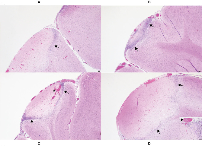
Figure 4.
Histologic images of affected cerebral gyri from the 5 s (A), 10 s (B), 15 s (C), and 20 s (D) applications. In all application times, the cerebrum subjacent to the horn bud contained areas of leptomeningeal and cortical necrosis, often rimmed by moderate to large numbers of gitter cells (arrows). Hemorrhage was also present in the 15 s application (C, asterisk). Occlusive thrombi were identified within leptomeningeal blood vessels of most applications, but most frequently in the 20 s application (D, inset, arrowhead). Hematoxylin and eosin. Scale bars = 100 microns.

