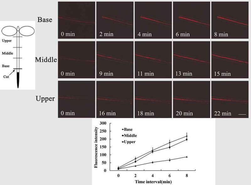
Figure 2.

Spatial–temporal variation of intracellular O2−· fluorescence in different parts of Arabidopsis wild-type (Col-0) hypocotyls cuttings. Wild-type seedlings were preloaded with DHE, then fluorescence images of intracellular O2−· were detected at the base, middle, and upper hypocotyl at different times after excision of primary roots. Data of fluorescence pixel intensities are displayed as means ±SE of three replicates, each replicate with 3 seedlings. Bar = 250 µm
