
Figure 4.
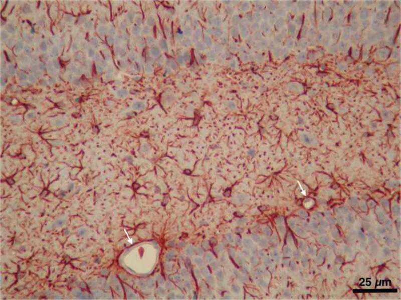
A light micrograph from our image archive showing astrocytes labeled by immunostaining for GFAP in the hippocampal region of the brain. Note the microvessels almost entirely surrounded with astrocyte endfeet (arrows)
Official websites use .gov
A
.gov website belongs to an official
government organization in the United States.
Secure .gov websites use HTTPS
A lock (
) or https:// means you've safely
connected to the .gov website. Share sensitive
information only on official, secure websites.
A light micrograph from our image archive showing astrocytes labeled by immunostaining for GFAP in the hippocampal region of the brain. Note the microvessels almost entirely surrounded with astrocyte endfeet (arrows)