
Figure 7.
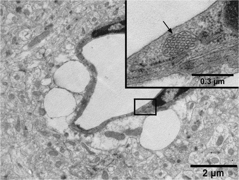
An electron micrograph from a previous study from our research group (reproduced from Ref. # 96 with permission from Elsevier Science) showing a capillary from the hippocampus region of the brain of a rat with cortical dysplasia exposed to febrile seizures. Note a conglomerate of caveolar vesicles within a cargo (arrow in the inset) in the cytoplasm of a brain capillary endothelial cell and the intensive pericapillary edema with swollen astrocyte endfeet