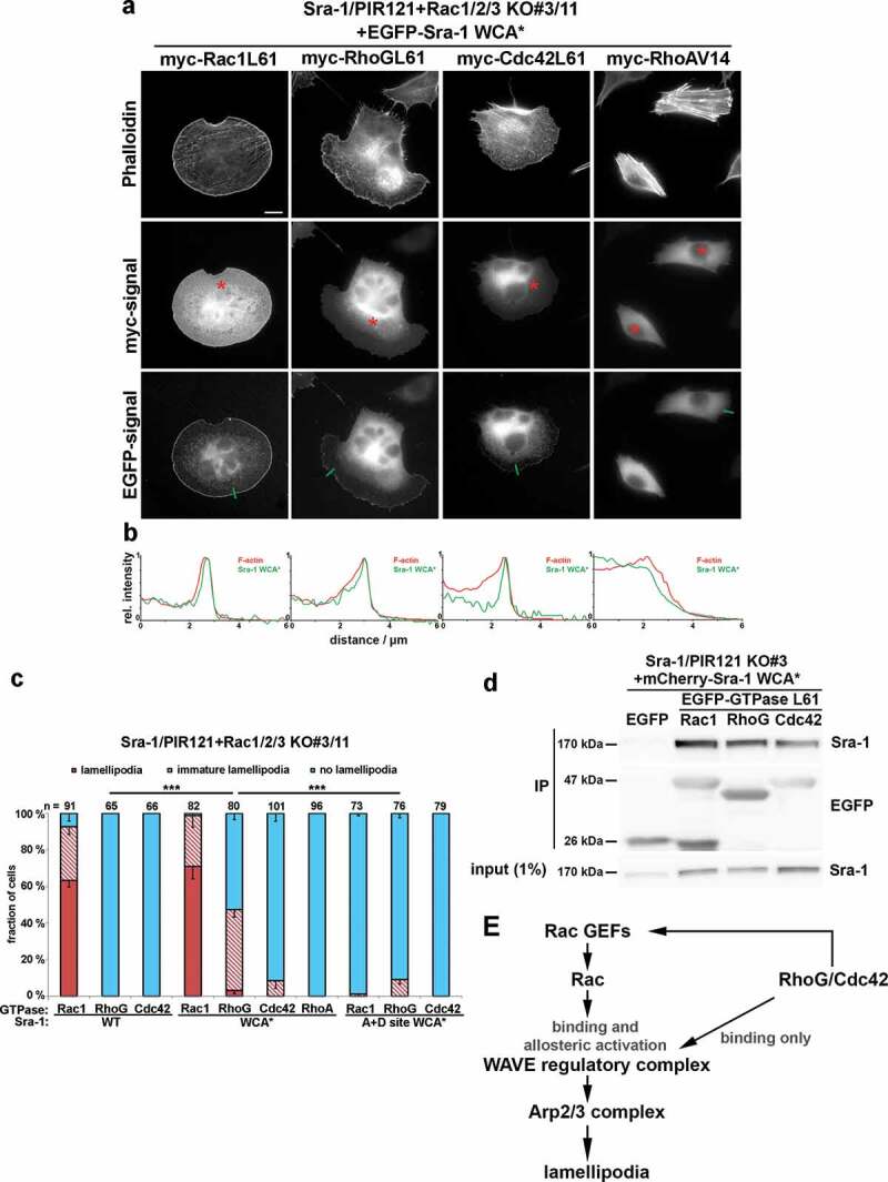
Figure 3.

Induction of lamellipodia by RhoG or Cdc42 in conjunction with activated WRC, but in the absence of Rac expression
(a) Representative images of Sra-1/PIR121+ Rac1/2/3 KO cells (clone #3/11) expressing EGFP-Sra-1 WCA* together with myc-tagged, small GTPases, as indicated. Transfected cells identified by myc-staining (asterisks) were analysed concerning their cell morphologies (top row, actin filament staining with phalloidin) and the capability to accumulate active WRC (Sra-1 WCA*) at peripheral lamellipodia (b). Measurements shown in b were performed along line scans as shown in the images provided in a (green lines). (c) Quantification of lamellipodial phenotypes, done as described for Figure 1(c). n gives number of cells analysed, and differentially coloured columns are arithmetic means ± SEM from at least three independent experiments. Statistical significance was assessed for differences between the percentages of cells with ‘no lamellipodia’ phenotype. ***p < 0.001 (two-sample, two-sided t-test). (d) Sra-1/PIR121 KO cells (clone # 3) were co-transfected with mCherry-Sra-1 WCA* and either EGFP or constitutively active (Q61L), EGFP-tagged small GTPases, lysed and subjected to immunoprecipitation against EGFP. Note the complete absence of immunoprecipitation of Sra-1 upon co-expression of EGFP alone (negative control), and significant interactions of Sra-1 WCA* with Rac-1, RhoG and Cdc42. (e) Model for how RhoG and Cdc42 might regulate WRC and lamellipodia formation.
