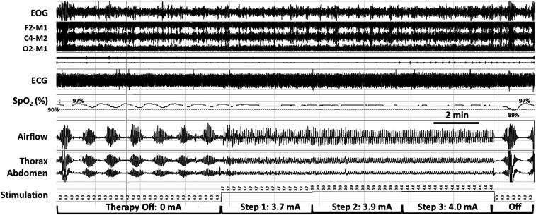
Figure 2. Automatic stepwise increases in stimulation amplitude stabilize tidal airflow, respiratory effort, and oxyhemoglobin saturation as ventilation is captured progressively.
A period of stepwise increases in phrenic nerve stimulation amplitude (middle segment) is bracketed by periods off stimulation that began with sudden movement arousals (just before the start of recording segment; far left) and at end of stable breathing period (see movement artifact in thorax and abdomen; right). Automatic stepwise increases in stimulation amplitude stabilize tidal airflow, respiratory effort, and oxyhemoglobin saturation as ventilation is captured progressively. Signals include EOG, EEG derivations as shown, ECG, SpO2 (pulse oximetry, %), airflow (nasal pressure cannula), and thoracic and abdominal piezoelectric girth sensors. ECG = electrocardiogram; EOG = electrooculogram; SpO2 = peripheral oxygen saturation.

