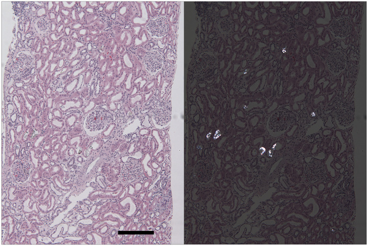
FIGURE 1.
CaOx deposition in a kidney allograft with delayed graft function at 20 days post-transplant. Left panel: this hematoxylin and eosin-stained section of kidney cortex reveals preserved kidney parenchyma with moderate distention of all tubules. Several tubules contain transparent crystals best visualized when the sections are examined under polarized light (right panel). Oxalate crystals are dissolved and disappear under the staining conditions of routinely used special stains, including PAS, Masson’s trichrome and Jones’ silver methenamine stains. (H&E; final magnification: 58×, bar = 250 µm).

