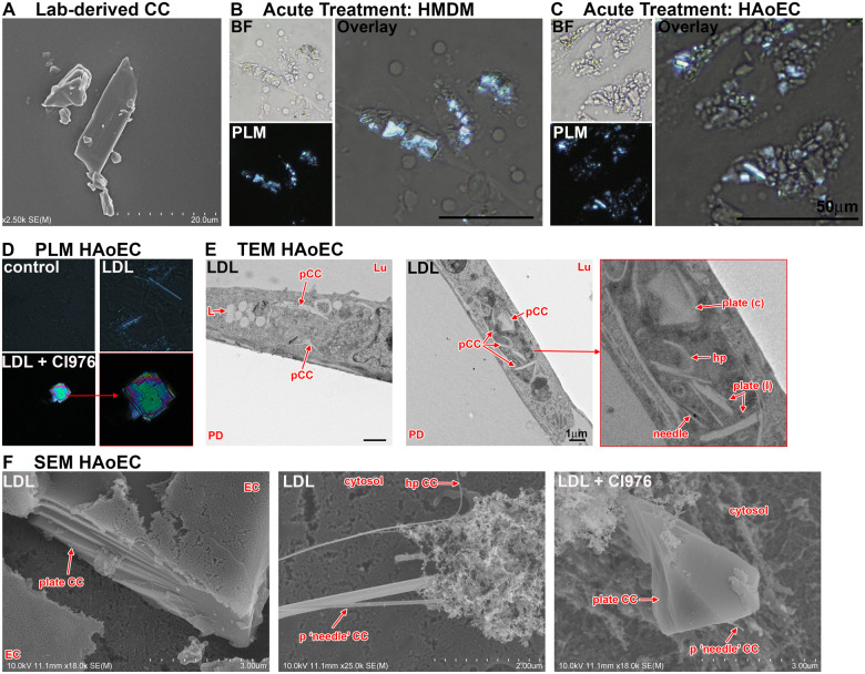
Figure 2.
Cholesterol crystals in macrophages and endothelial cells. (A) Lab-derived cholesterol crystals imaged by scanning electron microscopy. (B/C) Human monocyte derived macrophages (HMDM) and human aortic endothelial cells (HAoEC) were incubated with 100 μg/mL CC for 24 h, fixed using 4% paraformaldehyde fixation buffer and imaged by polarized light microscopy. Cholesterol crystal can be seen to be taken up by human monocyte derived macrophages and human aortic endothelial cells. (D–F) Low-density lipoprotein treated HAoEC where imaged using polarized light microscopy (D), transmission electron microscopy (E), and scanning electron microscopy (F). Various shapes, sizes, and orientations of cholesterol crystal can be seen by either technique. Importantly, the impact of lipid-altering substances like CI-976 on cholesterol crystal shape and size highlights the importance of future research into signalling pathways regulating cholesterol crystal formation, shape, size, and origin. BF, bright field; CC, cholesterol crystal; EC, endothelial cell; hp, hairpin shaped CC; L, lipid; Lu, lumen/apical cell side; pCC, possible CC; PD, petri dish; plate (C), plate-shaped CC cross sectioned; plate (l), plate-shaped CC longitudinal sectioned; PLM, polarized light microscopy.

