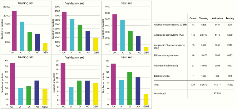
Fig. 2.
Six histological categories of glioma H&E sections were involved. 1) oligodendroglioma, 2) anaplastic oligodendroglioma, 3) astrocytoma, 4) anaplastic astrocytoma, 5) glioblastoma, and 6) nontumor images of red blood cells and background brain glia. The images were divided into 3 sets: 1) training set: 65 673 images from 219 subjects, 2) validation set: 14 317 images from 48 subjects, and 3) testing set: 16 862 images from 56 subjects. Bar charts for the number of patches and patients are displayed on the left, and detailed numbers of 5 subtypes for the training, validation and testing sets are shown on the right. Note that background image patches were taken from the boundary of tumor regions in the sections.

