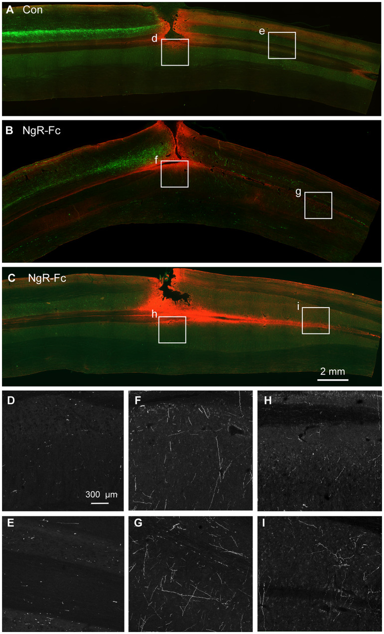
Figure 7.
NgR-Fc treatment increases growth of CST fibres caudal to the injury. Representative images of horizontal section of spinal cord stained for BDA (green) and anti-GFAP (red) from one vehicle-treated (A) and two NgR-Fc-treated animals (B and C). Rostral to the left and right side is up. The area in the white boxes from A–C (marked d–i) are magnified in D–I. Note numerous BDA-labelled CST fibres significantly increased in the NgR-Fc treated animals. Scale bar = 2000 µm in A–C, 300 µm in D–I.

