. 2021 Jan 8;3(1):e200006. doi: 10.1148/rycan.2021200006

Figure 2:

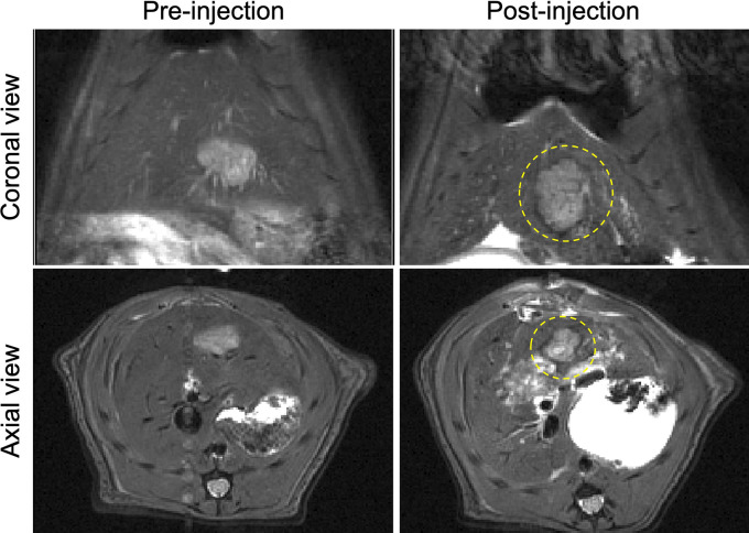
MR images obtained before and after the injection of sorafenib-eluting embolic microspheres (SOR-EMs) into the hepatic artery. Dotted circles outline regions of marked T2-weighted signal intensity changes caused by the deposition of iron oxide–labeled SOR-EMs at the typical hypervascular periphery of the tumor.

MR images obtained before and after the injection of sorafenib-eluting embolic microspheres (SOR-EMs) into the hepatic artery. Dotted circles outline regions of marked T2-weighted signal intensity changes caused by the deposition of iron oxide–labeled SOR-EMs at the typical hypervascular periphery of the tumor.