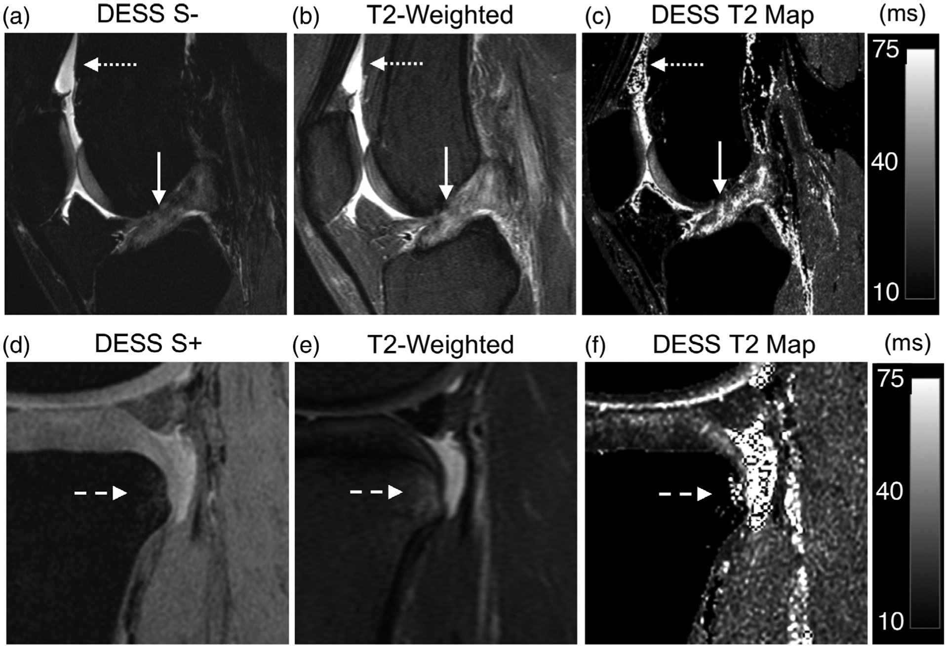
FIGURE 2:

A 19-year-old female presenting discontinuity of the anterior cruciate ligament (ACL) fibers, compatible with a complete tear (solid arrow) and mild joint effusion (dotted arrow). (a) A sagittal water-excitation DESS S− image demonstrates increased signal in the ACL and discontinuity of the ligament fibers as well high fluid contrast for the joint effusion. (b) A sagittal T2-weighted fat-saturated scan similarly demonstrates a full thickness tear of the ACL and increased signal intensity for the synovial fluid. (c) An instantly generated DESS T2 relaxation time map shows elevated T2 values of the ACL (similar to that of adjacent cartilage) and focal regions of fluid signals. (d) A bone marrow lesion (BML) in the same patient (dotted arrows) in the posterior tibial condyle is underestimated in volume by the DESS S + image. (e) A T2-weighted scan depicts the actual size of the BML. (f) The DESS T2 map shows the T2 of the BML with the same volume as the S + image.
