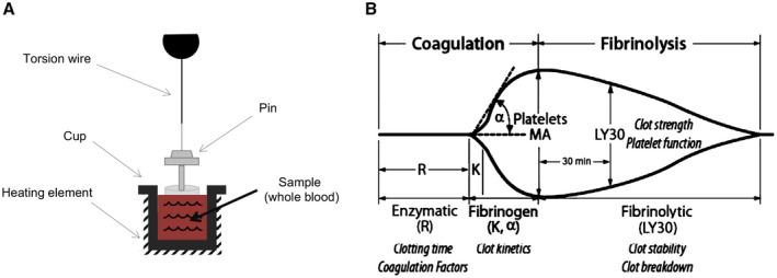
FIG. 1.

Thromboelastography. (A) TEG measures the properties of clot formation using a small cup that holds the blood sample and slowly oscillates. A pin held by a thin torsion wire is suspended in the blood; as clot forms, it binds the cup and pin together. The torsion on the pin is measured and converted to an electrical signal. Clot strength is directly proportional to torsion on the pin. (B) Graphical presentation of the TEG hemostasis profile for clot formation and lysis, with MA reflecting overall clot stability.
