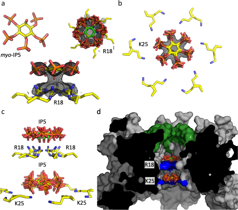
Fig 1. Structure of IP5 bound to HIV CA hexamer.
(a) (left) Structure of myo-IP5 ligand. Fo − Fc omit density (mesh) contoured at 2.0σ centered on IP5 bound to R18 and viewed down the 6-fold axis (right) and from the side (below). (b) Fo − Fc omit density (mesh) contoured at 2.0σ centered on a second IP5 molecule next to K25 (two rotamer side chains are shown). All six-symmetry equivalent IP5 molecules are shown. (c) View showing the two IP5 molecules, one binding above R18 and one above K25. Note the second IP5 molecule is located closer to the K25 ring than R18. (d) Cross section through the hexamer, showing the central chamber where the IP5 molecules are bound. The β-hairpin is shown in green and the location of R18 and K25 in blue.

