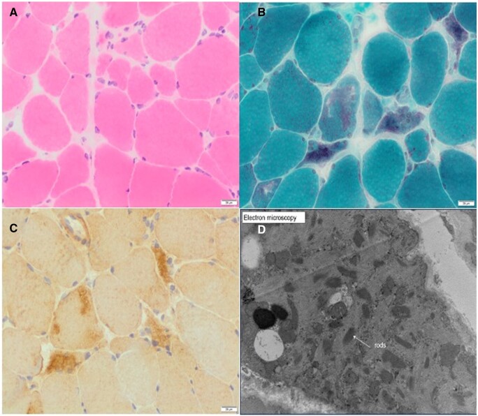
Figure 2.
Histopathological findings from left vastus lateralis biopsy. (A) H&E staining showing skeletal muscle with significant variability of muscle fibre diameters. Scattered atrophic fibres including round, polygonal, and angulated forms. There is no significant increase in endomysial connective tissue. A few rare pale necrotic fibres may be present. No obvious inflammation is present. (B) Modified Gomori trichrome highlights the presence of relatively frequently lobulated fibres and scattered fibres, which appear to contain aggregates of rod-like structures. (C) Myotilin staining showing myofibril aggregates, which co-localize with rods. (D) Electron microscopy confirms the presence of nemaline rods, which have a high electron density and an internal lattice structure.

