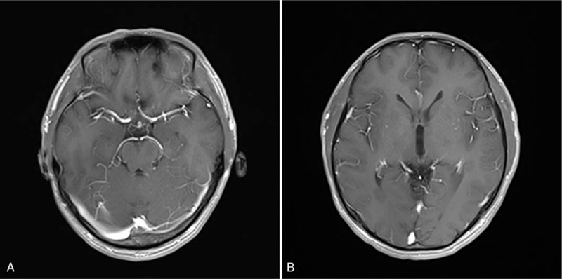
Figure 2.

Brain T1-weighted postcontrast MRI of anti-IgLON5 disease. Representative axial, T1-weighted postcontrast MRI demonstrating no significant postcontrast enhancement of left tegmentum of the midbrain (A) and right occipital horn of the lateral ventricle (B).
