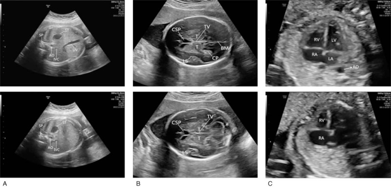
Figure 1.

Comparison of the standard plane (upper row) and nonstandard plane (lower row) in 3 sections. A, The lower abdominal FS image does not show the umbilical vein. B, The lower head FS image does not show the brain midline and the choroid plexus. C, The lower heart FS image does not show the left ventricle, left atrium, and descending aorta.
