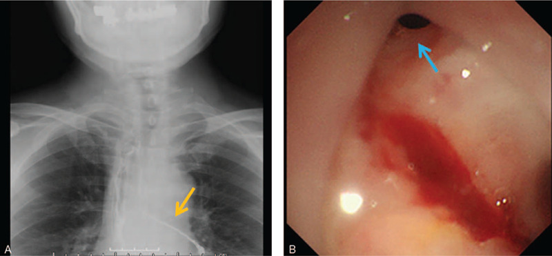
Figure 1.

The representative findings of esophago-bronchial fistula are shown. (A) Gastrografin contrast study shows leakage into the left bronchus (orange arrow). (B) A fistula is detected in the anterior wall of the esophagogastric anastomotic area by endoscopy (blue arrow).
