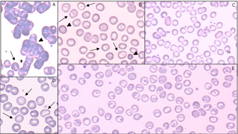
Figure 1. Blood smear features of five patients with COVID-19-related anaemia.
(A) A 40-year old man, on mechanical ventilation, at 43 days of hospitalisation. Blood type was A positive, direct antiglobulin test (DAT) positive, haemoglobin (Hb) 7 g/dL, mean corpuscular volume (MCV) 92 fl, red cell distribution width (RDW) 15.9% at the time of investigation. In this field, autoagglutination (arrowhead) and rouleaux (asterisk) formations are visible. At the bottom of the panel, stomatocytes (arrows) are visible. 80× objective. (B) A 47-year old man, on mechanical ventilation after 32 days of hospitalisation; under treatment with steroids, anakinra, quinolones. Blood type was A positive, DAT negative, Hb 11.3 g/dL, MCV 89 fl, RDW 12.4% at the time of investigation. Spherocytes (arrowheads) and knizocytes (arrows) are visible. 100× objective. (C) A 76-year old woman, on mechanically assisted ventilation, after 36 days of hospitalisation. Blood type was A positive, DAT positive, Hb 8.3g/dL, MCV 81 fl, RDW 14.6% at the time of investigation. This field is widely occupied by cup-shaped erythrocytes. Cytoplasmic rim is intensely coloured, surrounding a wider than normal central area. 100× objective. (D) A 66-year old woman, on mechanically assisted ventilation, after 49 days of hospitalisation, under treatment with hydroxychloroquine and piperacillin. Blood type was O positive, DAT positive, Hb 7.7 g/dL, MCV 88 fl, RDW 13.4% at the time of investigation. Stomatocytes (arrows) are present in this field. 100× objective. (E) A 69-year old man, on mechanical ventilation after 40 days of hospitalisation, under treatment with hydroxychloroquine, lopinavir, ritonavir. Blood type was A positive, DAT negative, Hb 7.4 g/dL, MCV 91 fl, RDW 15.4% at the time of investigation. In this field, red cells are totally represented by knizocytes 100× objective.

