Fig. 5.
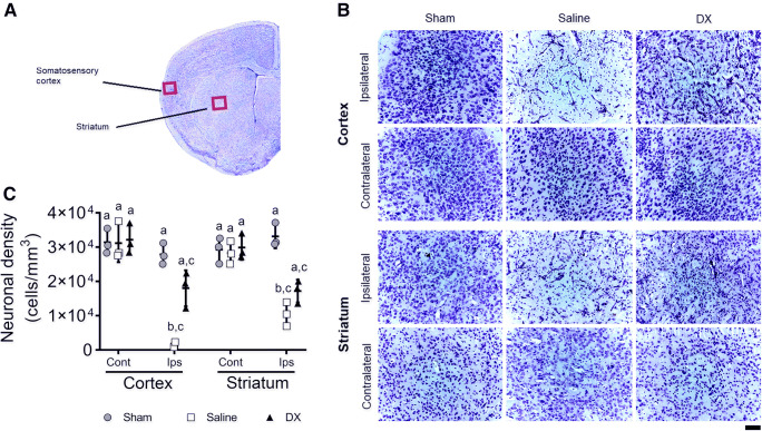
Increased neuronal density in the somatosensory cortex and the striatum after intranasal dexamethasone (DX) treatment in the MCAO model. Representative image of a brain section stained with Nissl to illustrate where neuronal quantifications were analyzed (A). Representative images of Nissl staining of the somatosensory cortex and striatum from the sham-, saline-, and DX-treated mice after 24 h of performing MCAO (B). Quantification of the neuronal density of 4 adjacent histological sections of 20 μm (800 μm total distance) stained with Nissl in the cortex and striatum from the ipsilateral (Ips) and contralateral (Cont) hemisphere of the sham, saline, or DX animals (C). Each point represents the data of an individual mouse (n = 3 per group). Data represent the mean ± SEM. Data were analyzed by a Kruskal–Wallis, followed by Dunn’s multiple-comparisons test. Different letters (a, b, and c) indicate significant differences in the neuronal density between groups (p < 0.05). Scale bar, 50 μm

