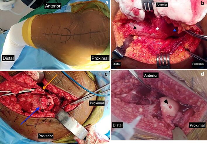
Fig. 1.
Intra-operative photographs of modified Dunn osteotomy performed on a left hip. a Incision centered over greater trochanter. b Deep exposure, just prior to digastric osteotomy; star—greater trochanter, black triangle—vastus lateralis, blue triangle—abductors. c Extensile retinacular flaps raised (yellow arrow), Dunn osteotomy being planned (blue arrow). d Slip reduced, active bleeding seen from epiphysis after reduction (arrow head)

