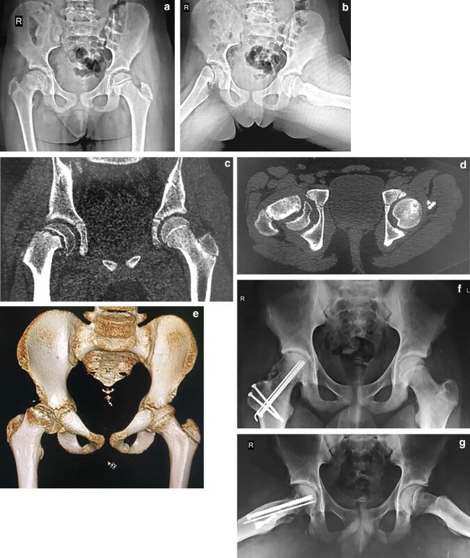
Fig. 2.
A thirteen-year-old boy with right-sided, acute on chronic, stable slip. Pre-operative X-ray pelvis with both hip joints, a anteroposterior and b frog leg lateral views showing a moderate slip (52°). c Coronal, d transverse, and e three-dimensional reconstruction computerized tomography images prior to surgery. f and g Radiographs at 2.5-year follow-up after capital realignment using modified Dunn procedure (final slip angle 3°, alpha angle 26°)

