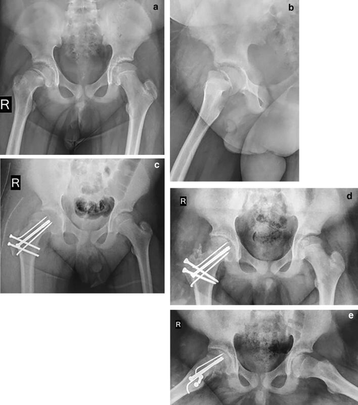
Fig. 3.
A thirteen-year-old boy operated for right-sided acute, unstable slip. X-ray pelvis with both hip joints a anteroposterior and b cross table lateral views at presentation. c Immediate post-operative anteroposterior radiograph showing subluxation. d Anteroposterior and e frog leg lateral radiographs at 18 months after revision surgery (removal of interposed capsular tissue and the trochanteric screws exchange). Note the concentric joint reduction and absence of AVN

