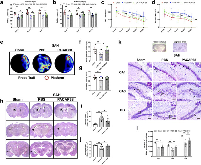
Fig. 6.
Exogenous PACAP38 treatment improved long-term neurological performance and prevented hydrocephalus and neuronal degeneration at 28 days after SAH. (a, b) Rotarod test (5 RPM and 10 RPM) on the first, second, and third weeks after SAH. (c, d) Escape latency and swimming distance of Morris water maze test on days 23 to 27 after SAH. (e) Representative thermal imaging images of the probe trial. (f, g) Quantification of probe quadrant duration and swimming velocities. (h–j) Representative images of coronary section at different levels, and quantification of ventricular volume (mm3) and cortical thickness (mm) in different groups. (k, l) Representative images and neuronal quantifications of Nissl staining in hippocampal CA1, CA3, and DG regions. Arrows indicate shrunken pyramidal or dead neurons. Scale bar = 200 μm. n = 10/group. *P < 0.05, **P < 0.01. One-way ANOVA, Tukey’s post hoc test for (f, g, i, j). Two-way ANOVA, Tukey’s post hoc test for (a–d, i)

