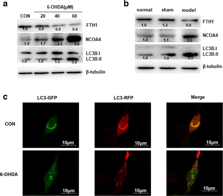
Fig. 9.
Induction of ferritinophagy in the 6-OHDA model of PD. The expression of NCOA4, LC3B-I, and LC3B-II in PC-12 cells (A) and SN tissue (B) from rats was determined by Western blotting. The data were normalized to the control group values. (C) PC-12 cells were infected with a GFP-RFP-LC3 adenovirus for 24 h and then treated with 40 μM 6-OHDA for 24 h. Autophagosomes, which are represented by yellow puncta in the merged images, and autolysosomes, which are represented by red puncta, were detected with fluorescence microscopy. The results represent the means of 3 independent experiments

