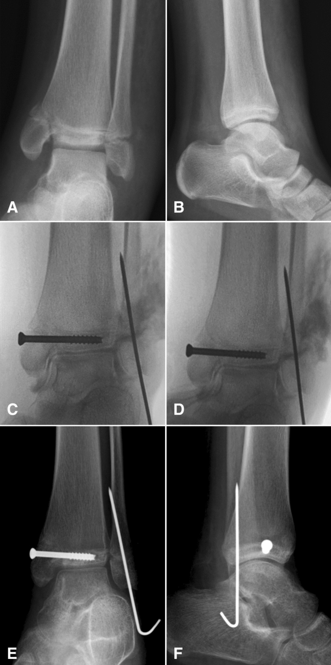
Fig. 6.

a, b AP & lateral X-rays of the ankle joint showing a SH Type IV injury of the distal tibia in a 10 yr old boy; c, d AP & Mortise views of the intra-operative C –arm images following closed reduction, fixation and arthrogram showing good reduction and articular congruity; e, f AP & Lateral X-rays done at 6 weeks follow-up showing good healing of the fracture
