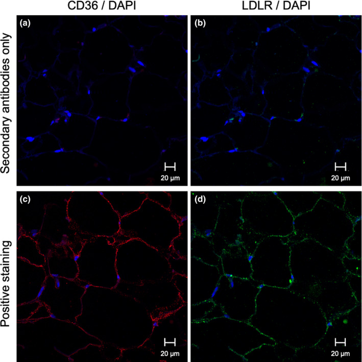
FIGURE 1.

Representative plasma membrane localization of CD36 and LDLR by immunohistofluorescence staining in WAT. Specificity of LDLR and CD36 detection in WAT was verified by secondary antibody‐only staining for CD36 (Alexa Fluor 555 anti‐rabbit IgG) (a), and LDLR (Alexa Fluor 647 anti‐rabbit IgG) (b), used as negative control; Positive staining (primary and secondary antibodies) of WAT section for CD36 (c) and LDLR (d) All samples counterstained with nuclear stain DAPI. A‐B represent the same WAT section, C‐D represent the same WAT section, all from the same subject
