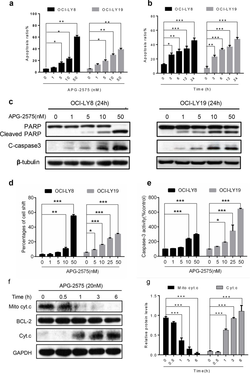
Figure 2.
APG-2575 induces apoptosis associated with mitochondria-dependent pathway. (a) OCI-LY8 and OCI-LY19 cells were treated with indicated concentrations of APG-2575 for 24 h and then detected apoptosis by annexin V/propidium iodide (PI) staining and analyzed using flow cytometry. (b) APG-2575 induced a time-dependent apoptosis in OCI-LY8 and OCI-LY19 cells determined by flow cytometry at 25 nM of APG-2575. (c) Western blot analysis to evaluate the protein expression of PARP/cleaved PARP and cleaved caspase 3 change in OCI-LY8 and OCI-LY19 cells treated with indicated concentrations of APG-2575 for 24 h. β-Tubulin was the loading control. (d) Mitochondrial membrane potential detected by JC-1 staining of OCI-LY8 and OCI-LY19 cells treated with indicated concentrations of APG-2575 for 24 h and quantification of the percentages of cells shift. (e) Caspase 3 activity was measured 24 h after adding APG-2575 and was plotted relative to the DMSO control. (f) Cytochrome c (Cyt. c) abundance in mitochondrial and cytosolic fractions of OCI-LY8 were determined by Western blot after 6 h of treatment with increasing concentrations of APG-2575. BCL-2 and GAPDH serve as protein loading controls for the mitochondria and cytosol, respectively. (g) Quantification analysis of Cyt. C relative expression in (f). The data are presented as the mean ± SD of triplicate experiments. Statistical significance was determined by one-way analysis of variance (ANOVA). *p < 0.05, **p < 0.01, ***p < 0.001.

