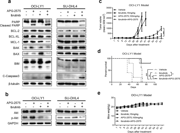
Figure 5.
Combination of APG-2575 and ibrutinib induces decreased expression of MCL-1 and p-AKT. (a) OCI-LY1 and SU-DHL4 cells were treated with DMSO, APG-2575 (10 nM for OCI-LY1, 50 nM for SU-DHL4), ibrutinib (1 μM), or both agents for 24 h. Expression of BCL-2 family members and apoptosis proteins were determined by Western blot. β-Tubulin was used as a loading control. (b) Western blot was performed to detect the expression of Akt and p-Akt in OCI-LY1 and SU-DHL4 cells treated with DMSO, APG-2575 (10 nM for OCI-LY1, 50 nM for SU-DHL4), ibrutinib (1 μM), or both agents for 24 h. (c) Tumor growth of OCI-LY1 model treated with vehicle, APG-2575 (100 mg/kg), ibrutinib (15 mg/kg), and Combo. Data are shown as mean ± SEM of five mice in each group. Statistical significance was determined by two-way ANOVA. ***p < 0.001. (d) Kaplan–Meier survival curves of mice between the four groups. n = 5 per group; statistical significance was evaluated by the log rank test. **p < 0.01, *p < 0.05, ***p < 0.001. (e) Mice weight of the four separate groups was recorded. Data are shown as mean ± SD of five mice in each group of triplicate experiments.

